Abnormal Microclots in Long Covid
Mechanism of Formation & Data from Long Covid Patients
Today I want to talk in more detail about the abnormal microclots that seem to be the cause of Long Covid and its associated symptoms.
Specifically, I want to walk through the evidence showing that these microclots exist in Long Covid patients and summarize what we know about their formation and how they are structured. This information is vital if we are ever going to find a way to stop these clots from forming and remove them from the bloodstream, which will hopefully treat and alleviate many of the symptoms of Long Covid.
How do Abnormal Microclots form in Long Covid?
Before we can talk about how abnormal microclots form in Long Covid, we have to talk about how clots form naturally in the healthy body.
In healthy people, we have a long, somewhat complicated process known as the “Coagulation Cascade” that involves over ten different enzymes and biochemical steps to initiate clotting. At the end of that cascade is one biochemical reaction that actually forms the clot: A monomeric protein called fibrinogen is modified by the enzyme thrombin in such a way that it polymerizes into what we call fibrin. This fibrin polymer is the structural foundation of what becomes a clot. Subsequent things happen to the fibrin polymer, like cross-linking to become stronger while maintaining flexibility and binding of platelets & other proteins, on its way to becoming a mature clot that will stop bleeding. When the body no longer needs the clot, we have an endogenous process called fibrinolysis that breaks the clot down. Fibrinolysis is a specific type of proteolysis that breaks down the protein structures by cleaving the amide bonds between certain amino acids.
So how do abnormal microclots form in Long Covid and how is that mechanism different from normal, healthy clot formation?
The Process:
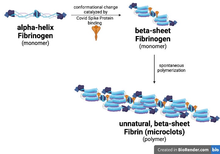
The formation of abnormal clots in Long Covid starts with the S1 subunit of the Covid Spike Protein binding to fibinogen. Binding of the Spike protein causes a conformational change in the fibrinogen monomer. The natural conformation of fibrinogen protein in a healthy body is mainly as alpha-helices. Binding of the Spike protein causes a change of the conformation of fibrinogen to mainly beta-sheets. This unnatural, beta-sheet conformation of fibrinogen polymerizes into fibrin ON IT’S OWN, without requiring thrombin enzyme as a catalyst, and forms the unnatural, abnormal microclots. The fibrin polymer in these abnormal clots is in the beta-sheet conformation. Spike protein is able to catalyze the formation of abnormal microclots at concentrations as low as 1 part per billion (1 ppb, Ref 1).
We typically refer to polymerized beta-sheet structures as amyloids. To give some context, amyloids that form in the brain are known to cause Alzheimer’s Disease (amyloids of A-beta protein), Parkinson’s Disease (amyloids of alpha-synuclein protein) and Creutzfeldt-Jakob Disease (amyloids of prion proteins). The abnormal microclots that form in Long Covid are amyloids of fibrin, again in this unnatural beta-sheet conformation.
Amyloids of beta-sheet conformation proteins also tend to be resistant to proteolysis and persist where they form. In the case of Long Covid microclots (amyloids of fibrin), this means that they persist in the vascular system and blood circulation.
The Data:
What data is available that shows that Covid Spike protein causes microclots?
In vitro data shows that if you add catalytic amounts (1 part per billion) of purified S1 Covid Spike protein to a sample of platelet poor plasma (PPP) from a healthy, Covid-naive person, abnormal amyloid microclots form. (Ref. 1) These clots can be detected by fluorescence microscopy — Panel A below shows untreated, healthy PPP and Panel B shows healthy PPP plus just Spike protein. The dye used, Thioflavin (ThT), specifically stains for amyloid proteins in the beta-sheet conformation, showing that these are NOT normal healthy clots in the alpha-helix conformation. Also, importantly, these clots form WITHOUT thrombin enzyme being present. Which means that Spike protein alone can cause formation of abnormal microclots, without needing activation of the Coagulation Cascade.
In a second in vitro experiment, the same investigators (Pretorius, Kell, and co-workers) fluorescent-labeled fibrinogen protein and examined clot formation under “normal” conditions by addition of thrombin enzyme (Panel A below) versus upon exposure to thrombin enzyme and catalytic amounts of Spike protein (Panel B below). (Ref. 1) The excessive fibrin clot formation in Panel B, as seen by the more intense green fluorescence, is caused by the Spike protein.
There is preliminary in vivo data showing that abnormal, amyloid microclots can be induced in a mouse model. (Ref. 2) But that data comes from a pre-print that has not been peer-reviewed, so I’m not going to discuss it directly. The pre-print is here if you want to review it for yourself. Feel free to contact me if you want to discuss it.
Do Long Covid Patients have Abnormal Microclots?
The short answer is YES.
Here’s The Data:
Pretorius, Kell, and co-workers examined platelet-poor plasma from a volunteer both before they contracted acute Covid infection and after they had developed Long Covid (See Panels A & B below). (Ref. 3) The PPP samples were simply stained with ThT to detect abnormal amyloid microclots and visualized by fluorescence microscopy. As you can see, all the samples from after the volunteer developed Long Covid contain abnormal amyloid microclots. And in all the papers examining microclots in Long Covid patients published from 2020 to 2023, abnormal amyloid microclots were detected in ALL Long Covid patients examined.
Where do we go from here?
Abnormal amyloid microclots clearly occur in Long Covid patients. They clearly form as a result of the Spike protein interacting with fibrinogen. Microclots clearly form independent of thrombin and the endogenous Coagulation Cascade, though it does appear that thrombin and other coagulation factors can increase the number of abnormal amyloid microclots that form. A lot of additional questions occur to me:
How do we keep these abnormal amyloid microclots from forming?
How does fibrinolysis break down these abnormal amyloid microclots?
What can we do to help people with Long Covid that likely have abnormal amyloid microclots?
Leave me a comment below and let me know what you think of this post.
Stay tuned for more posts that address the questions above.
And to follow my research, subscribe to my Substack!
References:
SARS-CoV-2 spike protein S1 induces fibrin(ogen) resistant to fibrinolysis: implications for microclot formation in COVID-19. Bioscience Reports 2021, 41, BSR20210611.
SARS-CoV-2 spike protein induces abnormal inflammatory blood clots neutralized by fibrin immunotherapy. bioRxiv preprint doi: https://doi.org/10.1101/2021.10.12.464152.
Persistent clotting protein pathology in Long COVID/Post-Acute Sequelae of COVID-19 (PASC) is accompanied by increased levels of antiplasmin. Cardiovasc. Diabetol. 2021, 20, 172.


So my lay understanding of what I read here is that the spike protein triggers the clotting process with the result that abnormal microclots are formed. So, because this is happening in Long Covid patients this seems to indicate that viral reservoirs are in fact in play? (Or is it that even dead Covid particles are also doing this in which case does this mean that the body can’t clear out the dead cells?) If there are viral reservoirs, the key is to get rid of them to stop the microclots. I have long covid and have just started doing high dose vitamin C IVs of 50 mg of Vit C per treatment. I am still extremely fatigued but my stamina has shown a very small improvement (likely not noticeable to anyone on the outside and has not greatly increased my functioning but I can have a shower without feeling like it’s then the only thing I can do that day.) The vitamin C is supposed to act as an antiviral and eliminate any viral reservoirs. It also has other benefits as an antioxidant and in healing wounds. (This does make me wonder about increased clot formation)
I have an alternate view on lung physiology that dismisses the notion of oxygen and carbon dioxide gaseous exchange
The article is titled
We breathe air not oxygen
I take you though all the steps that lead to this statement
Including how oxygen is manufactured
How oxygen is calibrated
Eg medical oxygen has 67parts per million of water contamination
Why oxygen is toxic, dehydrates and damages the alveoli
Lung physiology requires the air at the alveoli to reach 100% humidity
Can you see the problem?
The new take on lung physiology:
The lungs rehydrate the passing RBCs with iso tonic saline solution as they pass through the alveoli capillary beds
RBCs change from dark contracted dehydrated to plump bright hydrated form as they soak up the iso tonic saline solution the bursting alveoli bubbles throw upon the capillary sac
The airway mucosa conditions the breathe with salt and moisture
Find the article
Jane333.Substack.com